CT scan can fail. Delayed splenic hemorrhage is real.
- Carlos E Costa Almeida

- 29 de nov. de 2019
- 5 min de leitura
Atualizado: 7 de dez. de 2020
Delayed splenic hemorrhage (previous delayed splenic rupture) has been only reported in a few cases in worldwide literature. It is defined as a delayed splenic bleeding in a patient with a normal index image following a trauma. Delayed splenic rupture was first describe in 1920 by Dr. Baudet. The initial definition included a timeline. Delayed rupture was the one occurring 48h or more after a trauma, the “latent period of Baudet”. However, a review of several cases conducted 40y later, found that the 80% of delayed rupture occurred with 14 days, and there were cases occurring 70 days after the trauma event. In 1981 the first case in the CT era was reported and defined as a splenic bleeding occurring 48h or more after the trauma. Since then there have been some reports, and definition changed. Nowadays it is called delayed splenic hemorrhage, including delayed splenic bleeding in a patient with index image without splenic lesion following a trauma, occurring any time following the initial event.
Delayed splenic hemorrhage can occur any time following the initial event.
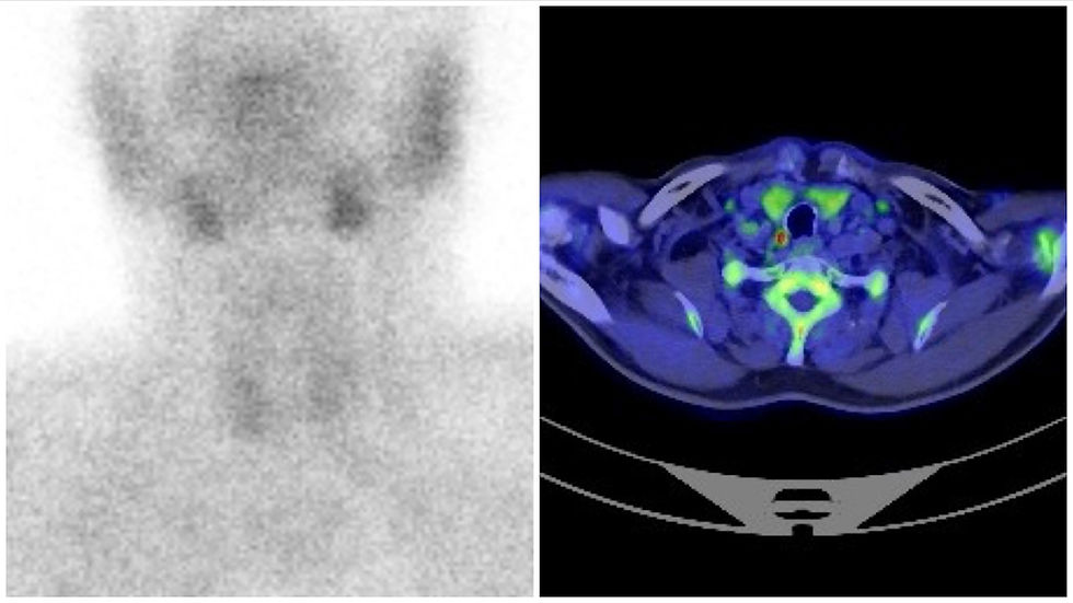
With the advent of new technologies and new CT scan devices, doctors do not know if this entity still exists or if it is only a myth in the present day. In this setting, a group of surgeons and radiologists from USA conducted a retrospective analysis of 6867 patients with splenic trauma in several institutions (Level I and Level II trauma centers – by definition in Portugal there is no Level 1 trauma center). Inclusion criteria: CT scan with intravenous injection performed at admission, with no splenic injury, no hemoperitoneum and no perisplenic hematoma. From those patients, 32 cases of delayed splenic hemorrhage (DSH) with eligible criteria were found.
DSH was diagnosed within the first 16 days following initial CT scan. Half of the cases were diagnosed in the initial 24h. Diagnosis was performed by new CT scan or intraoperatively. Hemodynamic instability and sudden drop of hemoglobin were the factors that lead doctors to the diagnosis. DSH occurred in all grade of splenic trauma (I to V from the AAST classification). The most common was grade I (47%), and the less common were grades IV and V (2% each). The authors reviewed the CT scans and took conclusions on their quality and technique used to diagnose splenic injuries. Results are interesting and should make us think. The majority of the initial CT scans (72%) were performed with an inadequate technique for splenic injuries diagnosis. Only eight patients were imaged in both portal and arterial phases. Additionally, only 9 scans were classified as good quality. Bad quality occurred because of artifacts due to arms placed adjacent to the torso.
Well, these data are very important. From them we can conclude that eventually some splenic injury might have been undiagnosed due to bad CT scans. Once again, surgeons/doctors must not blindly rely on image findings to decide patient management, I think. Clinical evaluation is crucial and the most important factor.
CT scans (72%) were performed with an inadequate technique.
From the 32 cases of DSH, 16 were submitted to some intervention. Fourteen (14) were treated by splenectomy (11) or splenorrhaphy (3). Two patients were managed by angio-embolization. The remaining 16 patients were submitted to a non-operative management. As it would be expected, grade I injuries were more likely to be treated by expectant management. One point I think is very important, is that two cases of splenic injury grade V (completely shattered spleen or hilar lesion that desvascularizes the spleen) were diagnosed as a DSH. Should we believe this type of lesion was not present at time of the first CT scan? Was the lesion missed in the initial CT scan? Is it possible to miss this type of severe lesion? If yes, how bad can a CT scan be to allow this to happen? Or, even a grade V splenic lesion can sometimes be evident only hours after the initial trauma and doctors must be aware of this. It seems that anything is possible when humans are involved. Once again, do not blindly rely on image findings.
The authors report two cases of angio-embolization to treat DSH. One in a grade III splenic injury, and one in a grade V splenic injury. This last one was a surprise to me. In fact, angioembolization can be of good help in cases of splenic hemorrhage in trauma patients, but in a grade V lesion I would never propose this approach. How can this work in a completely shattered spleen or in a hilar lesion that totally desvascularizes the spleen? According to the authors it can work. Do you agree? Would you adopt the same approach in a grade V splenic injury? Would you let a desvascularized spleen in a patient? Well… I think I would not. But we must be open minded.
CT scan must have portal and arterial phases.
According to Laura Harmon et al, a small subcapsular hematoma that was too small to be detected and then expanded and ruptured is a possible explanation for DSH. Subcapsular hematoma are in fact a risk factor for failure of non-operative management. Inadequate CT scan technique and/or bad quality CT scan are other possible reasons for a DSH. Only portal phase or only arterial phase CT scans can miss splenic injuries. Arms at the side of patient promote artifacts, and arms must be overhead (in CHUC-HG Covões in Coimbra, abdominal CT scans are perfomed with arms overhead). Following these possibilities for DSH etiology, authors give some advices for optimal CT scan technique in a trauma patient suspected of having a splenic injury: portal and arterial phase along with overhead arms are two of the four advices.
Surgeons/doctors must not blindly rely on image findings.
In conclusion, in the era of better CT scan devices DSH still exists and doctors must be aware of its possibility. One reason for DSH is a bad initial CT scan. In this setting, radiologists must know how to perform a good CT scan in the presence of an abdominal trauma patient who can have a splenic injury, and surgeons must know that CT scan and radiologists can also fail. CT scan is a great exam if it is well performed, but even so there is always a failure possibility. Always remember that serial clinical evaluation is of major importance to treatment decision, and that surgeons/doctors must not blindly rely on imaging findings.
Clinical evaluation always comes first!
Link to PubMed:
Dr. Carlos Eduardo Costa Almeida
General Surgeon


Comentários