Parathyroid cyst misdiagnosed with esophageal lesion. Success!
- Carlos E Costa Almeida

- 5 de ago. de 2024
- 4 min de leitura
Atualizado: 1 de nov. de 2024
Parathyroid cysts are extremely rare, represent <1% of all cystic cervical masses, and are responsible for less than 0.5% of parathyroid lesions. Women are more affected than men. Most parathyroid cysts are non-functional (80-90%), aspiration and PTH presence in the fluid is diagnostic, and treatment must be offered to all of them. They can be asymptomatic, can cause a mass effect compressing adjacent structures (causing dyspnea, dysphagia, hoarseness, cough, vein thrombosis), and can be responsible for 0.5-3% of primary hyperparathyroidism if functioning (10-20%).
According to Mohamed Amine Chaabouni et al. from Tunisia, these lesions can appear in the neck or the mediastinum (can cause RLN palsy), more frequently in the inferior parathyroids and on the left side. Surgeons must be aware that intrathyroidal parathyroid cysts have been described. The etiology of the parathyroid cyst is not well known, but degeneration of a parathyroid adenoma is a possibility. Misdiagnosis is a problem and can delay correct treatment. Mohamed Amine Chaabouni included as differential diagnosis thyroid cyst, parathyroid adenoma and carcinoma, branchial cleft cyst, vascular lesion, lipoma, adenopathy, neuroma, thymoma, lymphangioma, esophageal cyst or tumor, cystic hygroma, and mediastinal malignancy.
Recently, we had a female patient diagnosed with symptomatic primary hyperparathyroidism. Both neck ultrasound and SESTAMIBI were negative for parathyroid tissue. Bilateral neck exploration was not yet an option. There are more imaging exams available that can find the hyperfunctioning parathyroid tissue and allow a minimally invasive surgery (target parathyroidectomy).
A first neck CT scan showed a cystic lesion inferior to the right lobe of the thyroid, located in the upper mediastinum, posterior to the trachea and on the right side of it, pushing the esophagus to the left, with 48x29x22 mm, suggestive of necrotic adenopathy vs. esophageal duplication cyst. It is important to know, that a second CT scan raised the hypothesis of an enlarged parathyroid, and fine needle aspiration should be attempted (Figure 1).

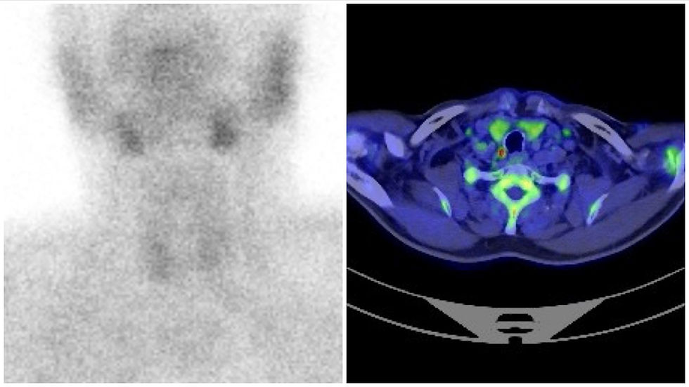
Meanwhile, an upper endoscopic ultrasonography was conducted, and the gastroenterologist suggested that the referred cystic lesion was an esophageal duplication cyst and that a fine needle aspiration would bring a high risk of infection. The surgical team decided to perform a PET-CT Choline. This exam showed a right-sided paratracheal nodule with peripheral uptake of the 11C-Choline (Figure 2).

Adding the information from the PET-CT Choline, the surgical team was convinced that the lesion was a functioning parathyroid cyst. A target right PIII parathyroidectomy was scheduled. Through an anterior approach, the referred cystic lesion was found deep within the inferior neck/upper mediastinum, inferior to the right lobe of the thyroid gland, adjacent to the trachea and esophagus, and posterior to the right carotid sheath. We were able to remove the entire cystic parathyroid without rupturing the cyst wall and preserving the right RLN. The cystic parathyroid was about 5 cm in length (Figure 3). Miami Criteria confirmed the success of the procedure:
Serum PTH before incision = 358.8
Serum PTH 0 min = 150.7
Serum PTH 10 min = 61.4
△PTH = 82.8%
Postoperative was uneventful, and the patient was discharged home on the next day. Pathology confirmed the diagnosis of a cystic parathyroid. Six months later patient was asymptomatic, with serum calcium and serum PTH within normal range.

Image diagnosis is challenging. Ultrasonography followed by FNA is usually the first technique. SESTAMIBI usually fails because the parathyroid tissue is compressed at the cyst peripheral and lacks tracer uptake, or there is dilution of the parathyroid tissue within the cyst fluid. SESTAMIBI has only 29% sensitivity for parathyroid cysts. That was the case of our case, with both ultrasound and SESTAMIBI negative for parathyroid hyperfunctioning tissue. CT and MRI will show a nonspecific lesion but can help surgeons evaluate the relationship with adjacent structures for correct surgical planning. In our case, PET-CT Choline was very useful to give us certainty about the preoperative diagnosis and promoting a target parathyroidectomy. Surgeons should use everything at their disposal so that the diagnosis is almost certain at the time of surgery.
FNA will show a water-clear fluid with high concentrations of PTH. The presence of PTH regardless of its level is suggestive of a parathyroid cyst. Additionally, FNA can be a first-line treatment option for nonfunctioning cysts. However, the recurrence rate is high and increases with cyst size. In case of recurrence surgery is the main option. Another treatment option after recurrence is sclerotherapy with tetracycline or ethanol. However, this technique is associated with leakage to the surrounding tissues, causing RLN injury and local inflammation. Surgeons must be aware that sclerotherapy increases the risk of complications if surgery is to be performed for recurrence. Surgery is always the first line of treatment for functioning parathyroid cysts, mediastinal parathyroid cysts, and suspicion of malignancy.
From these data, if the patient is suitable for surgery (no comorbidities), surgical resection should always be the first line of treatment, I think.
Link to reference
Dr. Carlos Eduardo Costa Almeida
General Surgeon


Comentários