Laparoscopic hernia repair: Injury to the corona mortis unleashes a demon.
- Carlos E Costa Almeida

- 9 de jul. de 2021
- 3 min de leitura
Minimally invasive inguinal hernia repair can be transabdominal preperitoneal approach (TAPP) or totally extraperitoneal approach (TEP). Laparoscopic repair is widely accepted because of less chronic postoperative pain, better cosmesis, and faster recovery comparing to open surgery. However, the laparoscopic approach can result in severe complications which are not common in open surgery, namely, deep infection (rare), visceral injury, and deep hemorrhage. Although deep hemorrhage can be life-threatening, it has a global incidence of 0,1%. A hemorrhage can be arterial or venous in its origin. Vascular injuries to the corona mortis, inferior epigastric vessels, external iliac vessels, obturator vessels, and iliac circumflex vessels can occur during TAPP or TEP.
Hemorrhage from the corona mortis can be fatal.
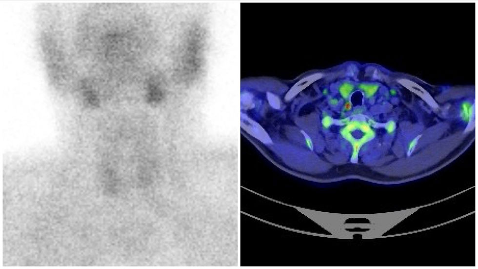
The corona mortis (picture) is a communicating vessel between the external iliac and obturator vessels. Its injury can result in massive bleeding and a postoperative hematoma needing another surgery. Tomohiko Yasuda et al. from the Nippon Medical School in Japan (Inzai and Tokyo) reported in 2017 one of these rare postoperative complications. During a TAPP repair, there was an apparent injury to the corona mortis with the electric hook, but no bleeding was noted during surgery. On postoperative day 1, swelling in the left lower abdomen suddenly appeared after the patient got out of bed. A CT scan confirmed a retropubic hematoma with active bleeding. An exploratory laparoscopy diagnosed bleeding from the corona mortis vein, and hemostasis was achieved with clips.

Hemorrhage from the corona mortis can be fatal. According to the authors, injury to the corona mortis artery can be diagnosed and treated by angiography. Injury to the corona mortis vein is a different issue and angiographic treatment is usually not easy. CT scan is the basis for diagnosing a corona mortis vein injury. First-line treatment is careful surveillance, and surgery is indicated only in cases of conservative management failure. Knowing the correct anatomy of the corona mortis is crucial to avoid this complication. The frequent location of the corona mortis is at the superior pubic ramus, with the distance to the pubic symphysis ranging from 21.4 to 91.0 mm, indicating a significant individual variation. This is important so that surgeons know that they should fix the prosthesis to the medial aspect of Cooper’s ligament avoiding the corona mortis vessels.
Bleeding from the corona mortis can be unnoticed during surgery.
Interesting from the case reported is the fact that no bleeding was noted during intraoperative injury. A possible reason is intra-abdominal pressure from the pneumoperitoneum. In fact, Tomohiko states that visibility of the corona mortis vessels can increase by reducing the pneumoperitoneum from 14 mmHg to 8 mmHg.
Should we decrease pressure if a vascular injury to the corona mortis is suspected? Can the pneumoperitoneum pressure help achieve definitive hemostasis?
Although both questions can have a positive answer, achieving definitive hemostasis by pneumoperitoneum pressure will eventually be possible only in low-pressure small vessels’ bleeding. Pneumoperitoneum can give us a wrong idea of definitive hemostasis, with bleeding still going on after CO2 evacuation. There is a take-home message from this case report: if a corona mortis injury is suspected and no bleeding is noted, surgeons should decrease pneumoperitoneum pressure to confirm hemostasis. It is easy to do, increases safety, and decreases surgeon’s anxiety.
If an injury to the corona mortis is suspected, decreasing pneumoperitoneum pressure can help confirm hemostasis.
Besides the intraoperative direct injury to the vessels, the authors include postoperative infection and mesh migration as possible causes of postoperative hemorrhage. There are reports of corona mortis hemorrhage following a slight migration of the mesh in the postoperative period with subsequent injury to the nearby vessels. In my opinion, this supports the idea of fixing the mesh and might be an advantage for self-fixating meshes usage. One final idea this report emphasizes is the use of laparoscopy to treat this complication. Image magnifying of anatomic structures is pointed out by the authors as the reason for the successful hemostasis with the laparoscopic approach.
Laparoscopic inguinal hernia repair has very good results with a very fast recovery. The authors conclude by stating that a “careful surgical manipulation and subsequent elaborate observation of the space around the corona mortis is critical for preventing delayed postoperative fatal hemorrhage.”
Link to PubMed:
Dr. Carlos Eduardo Costa Almeida
General Surgeon

Comentários