EGIST - a "GIST" outside the GI tract. New publication.
- Carlos E Costa Almeida

- 14 de dez. de 2019
- 3 min de leitura
Atualizado: 7 de dez. de 2020
Gastrointestinal stromal tumour (GIST) arises from the interstitial cells of Cajal in the intestinal wall. GIST can present as an exophytic mass projecting into the lumen or to the peritoneal cavity. However, a similar tumour can occur outside the gastrointestinal track with histopathological and molecular characteristics identical to the GIST. It is called extragastrointestinal stromal tumour (EGIST). Reith from USA was the first to use this name in 2000. Its origin is not fully known. Some authors state that it arises from Cajal-like cells outside the GI wall, while others believe it arises from pluripotent stem cells outside the GI track. Although EGIST is a very rare entity, it can occur as an abdominal mass hard to diagnose. Doctors must know it is a possibility so that an appropriate treatment can be offer to the patient.
EGIST is very rare and aggressive.
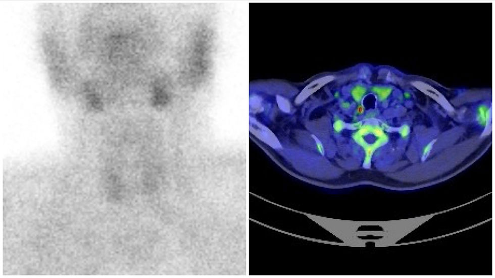
In that setting, I would like to share a recent publication in the BMJ Case Reports about an EGIST that me and my colleagues treated in the Surgery Department of the CHUC-Hospital Geral (Covões) in Coimbra, Portugal. A male patient resorted to the emergency department due to asthenia and melena, with severe anemia (4,3 g/dL). Following an upper gastrointestinal endoscopy and colonoscopy not conclusive for a diagnosis, an abdominal CT scan was performed. A huge tumour mass of 21 cm was found suggestive of a mesentery or exophytic tumour from the small bowel. A core biopsy was conducted concluding for a mesenchymal-like neoplasm without CD-117, desmin, S100, beta-catenin. Most probable diagnosis was a small bowel GIST.

Due to the severe anemia resistant to red blood cell transfusions and iron supplementation, we decided to perform a laparotomy to attempt complete tumour resection or tumour debulking. During surgery a tumour mass in the small bowel mesentery and the retroperitoneum, with encasement of the superior mesentery vessels and compression of the inferior vena cava was found. It was not resectable. Additionally, an omental-cake with small whitish haemorrhagic nodules and bloody peritoneal fluid was found. Omental resection was conducted to achieve the most tumour debulk possible, aiming at symptoms control and increase postoperative imatinib response. Pathology revealed an EGIST with high mitotic index (>5 mitoses/50 HPF), disseminated, and positive for c-KIT (CD117), DOG1 and CD34. Unfortunately, there was a fast decline in patient’s clinical status, who died two weeks following surgery.
Immunohistochemistry is crucial for diagnosis.
EGIST is a very rare and aggressive tumour. There are only 60 and 114 case reports of EGISTs in the retroperitoneum and mesentery, respectively. It represents about 1% of all gastrointestinal malignancies. Because of its rarity, imaging findings are not usually able to make a diagnosis. In fact, preoperative diagnosis is extremely rare. Although histology is similar to GIST, there are some differences:
While GIST usually presents with gastrointestinal hemorrhage and/or fistula, EGIST is asymptomatic in most cases. According to some authors, the most frequent presentation of an EGIST is abdominal pain followed by a palpable abdominal mass.
While GIST can affect any part of the gastrointestinal tract, EGIST can be located to the mesentery, retroperitoneum, omentum, pancreas, liver, gallbladder, urinary bladder, pleura, pelvis, vagina, prostate.
While GIST does not present with lymph nodes enlargement, EGIST usually does.
For EGIST diagnosis histology and immunohistochemistry are the gold standard. A positivity for CD117 (c-KIT) is necessary for confirmation (100%). However, other biomarkers are also useful for diagnosis, like positivity for CD34 and DOG1 presented in our case. While tumour size and mitotic count dictate the grading and prognosis of GIST, these are not yet valid in EGIST, although there are some authors who use in EGIST the same approach used for GIST. The only data available is that EGIST is more aggressive and has a worse prognosis, with an overall 5-year survival of 48,9%, contrasting with the 82,4% for the GIST.
Surgery is the most frequent approach. Imatinib remains uncertain.
The most usual approach to treat EGIST is surgery, aiming at debulking tumour whenever possible along with neighbor infiltrated structures. The utility and efficacy of imatinib mesylate remain uncertain due to the different behavior of EGIST compared with GIST. KIT mutation at exon 11 is associated with a good response to imatinib, and this mutation was only found in 37,5% of EGIST, while it is present in most GIST cases. Even though, imatinib use is advised for unresectable tumours.
As you can understand, a low threshold of suspicion is crucial, and EGIST must be included in the differential diagnosis of an abdominal mass. The absence of guidelines for EGIST treatment can drive some doctors crazy due to the excessive and pathological guidelines’ dependence of nowadays medicine. We need "raw medicine" to treat EGIST. There is "no net", there is only science and art.
Link to article:
Costa Almeida C, Caroço TV, Albano M, Carvalho L. Extragastrointestinal stromal tumour (EGIST) presented as a mesenteric and retroperitoneal mass. BMJ Case Rep 2019; 12:e2324871.
Dr. Carlos Eduardo Costa Almeida
General Surgeon


Comentários