Cannot find the parathyroid adenoma? Do not operate without a PET-Choline.
- Carlos E Costa Almeida

- 19 de out. de 2023
- 4 min de leitura
About 85% of primary hyperparathyroidism (PHPT) is caused by a solitary adenoma. Multiglandular disease and parathyroid carcinoma are responsible for 15% and 1% of cases, respectively. After the clinical and biochemical diagnosis of PHPT, the affected gland should be identified before surgery. Why? Successful preoperative localization of the diseased parathyroid gland (or glands) is mandatory for a minimally invasive approach. Nowadays, bilateral cervical exploration is not the rule and should be avoided (due to its increased morbidity). A minimally invasive surgery is the rule, meaning the surgeon directs the dissection towards the parathyroid gland he knows to be affected in advance.
According to Meghana Prablu et al. from New Delhi (India), the two main reasons for a failed surgery are “ectopic glands and undetected multiglandular disease”. So, for targeted parathyroid surgery (minimally invasive surgery) doctors need to use sensitive and accurate imaging techniques. To promote targeted and minimally invasive parathyroid surgery, surgeons need to have two concordant preoperative imaging tests showing the diseased parathyroid gland. Ultrasonography and scintigraphy are the most frequently used, identifying most parathyroid adenomas. However, 99m Tc-Sestamibi or MIBI (the most common tracer used) has a sensitivity of up to 89%. One-third of adenomas are invisible to MIBI. There are several disadvantages of this imaging modality:
Low sensitivity in small parathyroid adenomas due to its low spatial resolution
Adenomas near the thyroid
Multiglandular disease
Sometimes, using ultrasonography and MIBI no adenoma nor hyperplasic glands are found. In that setting, another option is available before promoting a bilateral cervical exploration. PET/CT with 18F-flurocholine (18F-FCH PET/CT) – PET-Choline – is recently being used for parathyroid adenoma identification and localization. PET-Choline offers a better spatial resolution than the remaining nuclear medicine imaging tests and offers a correlation of functional and anatomical information. Additionally, PET-Choline can identify smaller adenomas and reduce scanning time. Should it become the standard of care? Do not know…
There are several data in worldwide literature supporting the use of PET-Choline. Some authors reported that 18F-FCH PET/CT is not only useful in patients with a negative sestamibi scan but may also identify other parathyroid lesions in patients who were already submitted to MIBI. PET-Choline is particularly useful in patients with multiple lesions or hyperplasia, having better results than conventional imaging modalities. PET-Choline is also better for the identification of “smaller lesions which are very close to the thyroid”. According to the authors, PET-Choline has a sensitivity of 92% and specificity of 100% and can solve “discrepant results between MIBI scan and ultrasonography”.
Additionally, some concluded that with the advent of PET-Choline for target parathyroidectomy, there was no need for ioPTH testing. Well… for this I have some concerns. Using ioPTH testing and Miami criteria helps surgeons confirm the success of the minimally invasive surgery. It only takes 3-4 peripheral blood samples and 10 minutes. Why should we stop doing it?
Very important is the report of patients with negative MIBI and ultrasonography, who were submitted to minimally invasive parathyroid surgery after a positive 18F-FCH PET/CT. I believe this data is critical because a targeted surgery was possible instead of a bilateral cervical exploration with its associated increased morbidity. PET-Choline made it possible to decrease the risk of complications. Always remember that no surgery is free of risks, and doctors must use all available tools to preoperatively reduce those risks.
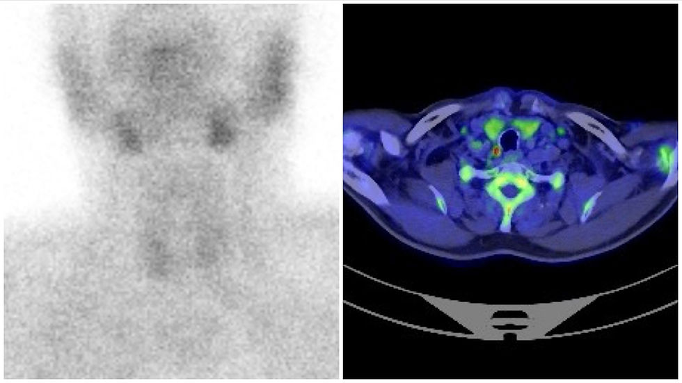
There are other PET tracers available. One of them is the 11C-Choline which has a high specificity for detecting parathyroid adenomas. The following images present a recent case I had in the Head and Neck Surgery Department of the Portuguese Oncology Institute of Coimbra (Portugal), of a male patient with negative ultrasonography and MIBI, but with positive 11C-Choline PET/CT for a PIII adenoma. This is a case where PET-Choline avoided a bilateral cervical exploration and promoted a targeted surgery. This is the way, I think.

Meghana Prablu et al also report a comparison between 4D-CT and PET-Choline. Both exams have 100% concordance in the localization of parathyroid lesions, but 4D-CT has a higher radiation dose. A polar feeding vessel, the “polar vessel sign”, may be found in the 4D-CT scan in up to 63% of adenomas. This sign increases the accuracy of the exam when present. 4D-MRI is another possibility, with higher sensitivity and specificity than 4D-CT and with no radiation, but with more “artifacts related to motion and swallowing”. However, there is a lack of reports at present in worldwide literature. Another recent possibility is PET/MRI. 18F-FCH PET/MR may have good results with less radiation exposure, no risk of contrast allergy, and better resolution. Reports are also lacking.
Finally, for secondary hyperparathyroidism, there are not many studies. We know that MIBI has a low sensitivity for hyperplasic glands. PET/CT looks better for multiglandular disease and MEN syndromic patients, though its significance is yet to be established.
In sum, PET-Choline is a very useful tool to help surgeons identify the affected gland and promote a targeted surgery to treat a PHPT due to an adenoma. According to the authors, the superiority of PET-Choline over MIBI has been proven. Should it become the standard of care? Probably, but until then PET-Choline should be used in cases of negative ultrasonography and MIBI, or cases of non-concordant imaging findings. This is the way to offer the patients the best surgical strategy, I think.
Link to article: Prabhu M, Damle NA. Fluorocholine PET Imaging of Parathyroid Disease. Indian Journal of Endocrinology and Metabolism 2018; 535-541.
Dr. Carlos Eduardo Costa Almeida
General Surgeon
#parathyroid #hyperparthyroidism #adenoma #parathyroid_adenoma #PET #Choline #18F #FCH #11C #targeted_surgery #minimallyinvasive #endocrine #endocrinology #CT #MRI #fluorocholine


Comentários