Autofluorescence in the treatment of hyperparathyroidism. HQ pictures.
- Carlos E Costa Almeida

- 29 de mar. de 2022
- 3 min de leitura
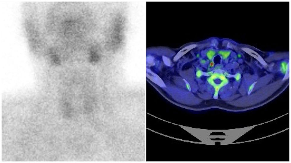
Parathyroid adenoma is a common cause of primary hyperparathyroidism. When surgical resection is indicated, pre-operative imaging findings will tell us which parathyroid is the diseased one, and where it is located. Ultrasonography, CT scan, and sestamibi scintigraphy are all used to diagnose and locate the parathyroid adenoma. Ideally, at least two of them should diagnose the adenoma in the same gland. This preoperative evaluation is paramount for successful surgical resection. However, sometimes imaging findings are not concordant and raise doubts about where the adenoma truly is.
Autofluorescence is a useful tool for parathyroids identification. When stimulated by near-infrared light, the parathyroids emit autofluorescence. This feature can be very useful in parathyroid and thyroid surgery since parathyroid identification during surgery can be very challenging. When stimulated by a laser beam of 795 nm, the parathyroid glands emit autofluorescence capable of distinguishing them from other tissues in the area. This is also true for an excised parathyroid. A major drawback of autofluorescence is the inability to differentiate normal parathyroid tissue from parathyroid adenoma or hyperplasia. In that setting, gland appearance and the surgeon’s experience will still have a major role in difficult cases.
An interesting paper from 2019 published by my friend Dr. Carlos Serra in BMC Surgery, reports the use of autofluorescence in patients with primary hyperparathyroidism due to adenoma with non-concordant preoperative imaging findings (ultrasonography and sestamibi scintigraphy). The author identified the glands with autofluorescence and removed the gland with the appearance of adenoma. Miami Criteria were used to confirm the cure. According to Carlos Serra et al., the autofluorescence intensity of the parathyroid is almost 2 times higher than the thyroid. Additionally, there was no difference in autofluorescence intensity between normal and diseased parathyroid glands. From the cases presented by Dr. Carlos Serra, I would like to highlight two of them. In both cases, PIII was preoperatively diagnosed as a diseased gland. The diagnosis was made by ultrasonography (sestamibi was negative) in one patient, and in the other, the diagnosis was made by sestamibi scintigraphy (ultrasonography was negative). During surgery, autofluorescence allowed the surgical team to find that the adenoma was in a PIV gland and not in a PIII. So, autofluorescence changed the gland to be removed, and was crucial to achieving a cure.
I had recently the opportunity to use autofluorescence in IPO Coimbra (Head and Neck Surgery Department) to treat primary hyperparathyroidism due to a right PIII adenoma diagnosed by ultrasonography and sestamibi scintigraphy. I used the "EleVision - IR Head & Neck" system from Medtronic. Preoperative imaging findings located the adenoma adherent to the posterior thyroid parenchyma of the right inferior pole (type A). PTH was measured three times: before skin incision, immediately before adenoma excision (0-min), and 10 min after resection. I used a “back door” approach (learned from my friend Prof Jaime Vilaça), and the suspected gland with adenoma was visually identified. Autofluorescence "EleVision" system confirmed and outlined the parathyroid tissue adherent to the posterior thyroid parenchyma. A right PIV was also identified and left in place. Miami Criteria proved the success of the procedure.
Despite the fact this was not a difficult case, my intention with this post is to share with you two beautiful pictures from this parathyroid surgery through a “back door” approach (Fig. 1), and the incredible image the autofluorescence can give you (Fig. 2 - Gif). Figures by CE Costa Almeida.


Some would say autofluorescence should only be used in selected cases (e.g., non-concordant preoperative imaging findings). However, an easy case can suddenly become a very difficult one. So, in my opinion, all adjuncts should be used to increase the surgical success rate and patient safety. Autofluorescence should also be used in total thyroidectomies to increase parathyroid preservation and decrease permanent postoperative hypoparathyroidism.
Attention to detail is the highway to success.
Link to article:
Dr. Carlos Eduardo Costa Almeida
General Surgeon
#parathyroid #adenoma #parathyroid_adenoma #autofluorescence #hyperparathyroidism #primary_hyperparathyroidism #thyroid #endocrine_surgery #parathyroid_surgery #thrydoid_surgery #total_thyroidectomy

Comentários